Everyone has at least one mole on the skin. However, not all moles are the same. Depending on the type, moles vary in shapes, and learning to distinguish them is essential for some of them require early diagnosis. Hence, let’s examine not only the characteristics and treatment methods of the various moles (birthmarks and beauty spots) that can occur on our skin but also melanoma, a malignant tumor of the skin that can be easily mistaken as a beauty spot. <Editor’s Note>
Congenital melanocytic nevus and dysplastic nevus can develop into melanoma, a malignant tumor. Melanoma is a type of cancer originating from melanocytes, cells that produce melanin pigment. It usually develops due to genetic factors or environmental factors such as exposure to ultraviolet radiation, especially appearing more commonly on Caucasians who lack melanocytes.
However, according to the National Cancer Center, although it is lower than that of the US or Australia, the incidence rate of melanoma cancer for South Korea is around 1 per 100,000 population. Also, although melanoma develops on the skin, early discovery and treatment are important since it can spread to the eyes, ears, and the mucous membrane of reproductive organs.
■The characteristics of congenital melanocytic nevus and dysplastic nevus
Congenital melanocytic nevus, which exists from birth, is likely to become thicker and grow hair as age. This nevus is classified by the predicted size at adulthood: ▲below 1.5cm is a small size, ▲1.5-20cm is a medium-size ▲bigger than 20cm is a large size. The growth of the head is 1.7 times, the body and arms are 2.8 times, and the legs are 3.3 times compared to at birth. In other words, if there is a nevus of a size bigger than 12cm on the head, 7cm on the body or the arms, 6cm on the legs since birth, then the nevus is predicted to become a larger size.
For dysplastic nevus, it is usually of a size larger than 6mm with irregular boundaries and with dark brown, black, and pink colors. Both congenital melanocytic nevus and dysplastic nevus have the potential to develop into a melanoma.

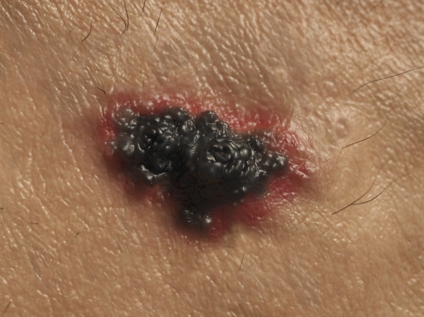
■When should we suspect melanoma?
Even if nevi develop into melanomas, most of them have no observable symptoms like pain. Therefore, the lesion needs to be observed with the naked eye to check if it develops into a melanoma. Here, the “ABCD rule” can be useful. This rule stands for ▲Asymmetry, ▲Border Irregularity, ▲Color variegation, and ▲Diameter. In other words, if the mole is larger than 0.6cm, is asymmetric, has irregular borders, and varies in color, you can suspect melanoma.
Furthermore, if bleeding, pain, or itching occurs, you need to visit the hospital.
■Regular observation and diagnosis needed
The bigger the congenital melanocytic nevus grows, the higher the possibility of malignant melanoma. Especially for large size nevus bigger than 20 cm, the possibility is 6-11%. Hence, it is better to get rid of a large size nevus in an early stage. In the case of nevus smaller than medium size, periodic observation is necessary, although complete removal is unnecessary due to the low possibility of developing into malignant melanoma.
Removal of dysplastic nevus is only recommended if there is a high malignant risk according to doctors’ diagnosis or difficulty in continual monitoring. Because melanoma develops even on the normal parts of the skin for individuals with a dysplastic nevus, periodically receiving skin checkups is of necessity even after the removal of all nevi.
“Even after surgery, the remaining cells at the boundaries of the removal site can grow and relapse into a nevus. There is no other way to prevent recurrence besides removing the nevus with sufficient space and expanded boundaries. Removing a large part of the skin is often difficult, depending on the size and location of the nevus. Therefore, a realistic solution would be regularly observing the surgical area even after surgery and receiving surgery once the nevus reoccurs before it’s too late,” Professor Na Jung-im from the Seoul National University Bundang Hospital commented.